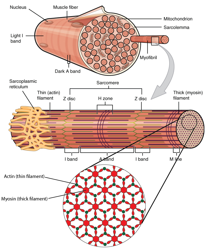
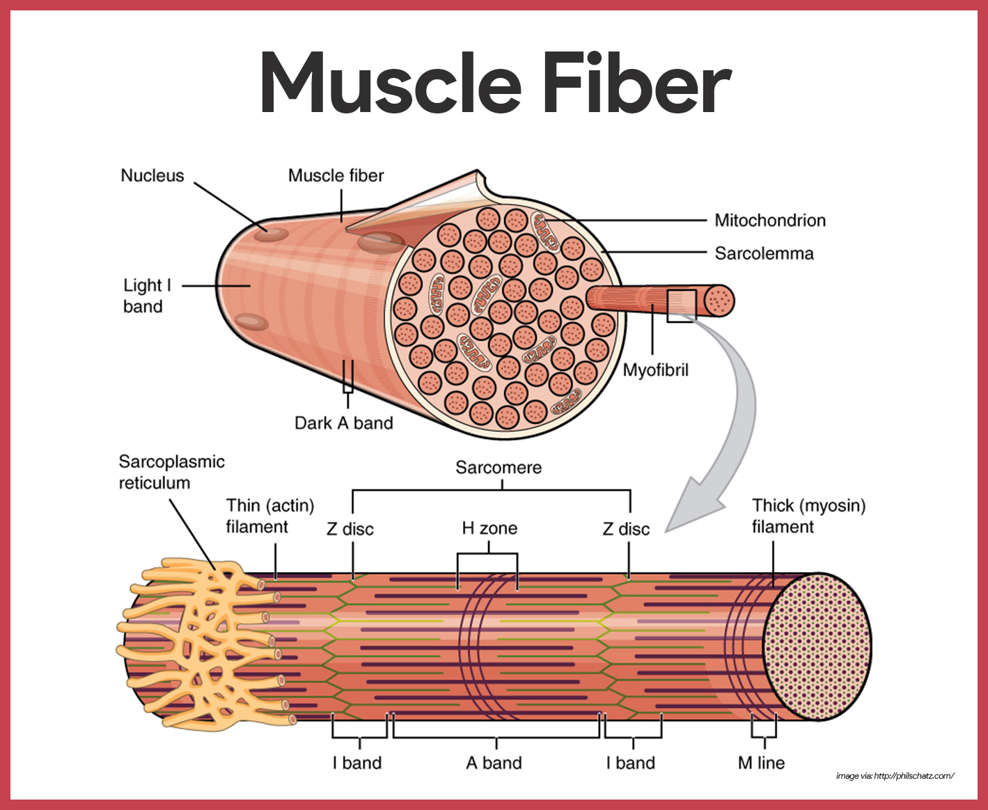
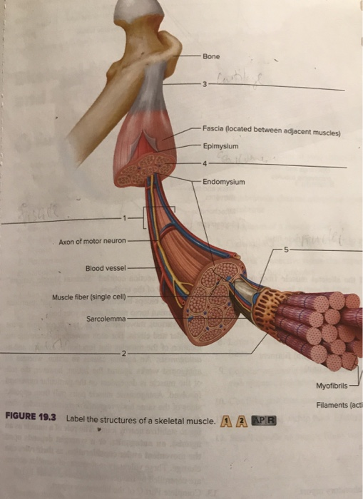
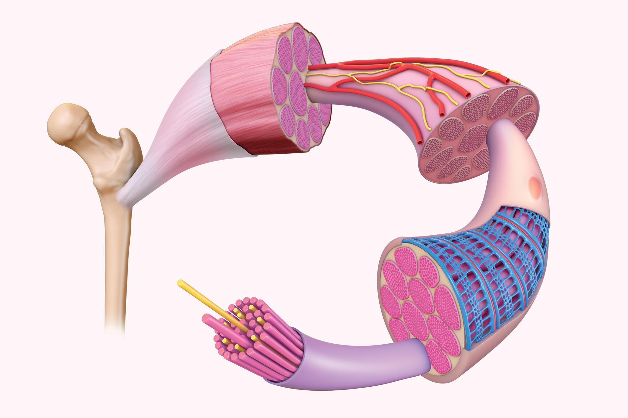
43 label the structure of the muscle fiber
Label structure of skeletal muscle Diagram | Quizlet Start studying Label structure of skeletal muscle. Learn vocabulary, terms, and more with flashcards, games, and other study tools. Neuromuscular junction: Parts, structure and steps | Kenhub At its simplest, the neuromuscular junction is a type of synapse where neuronal signals from the brain or spinal cord interact with skeletal muscle fibers, causing them to contract. The activation of many muscle fibers together causes muscles to contract, which in turn can produce movement. The neuromuscular junction then, is a key component in ...
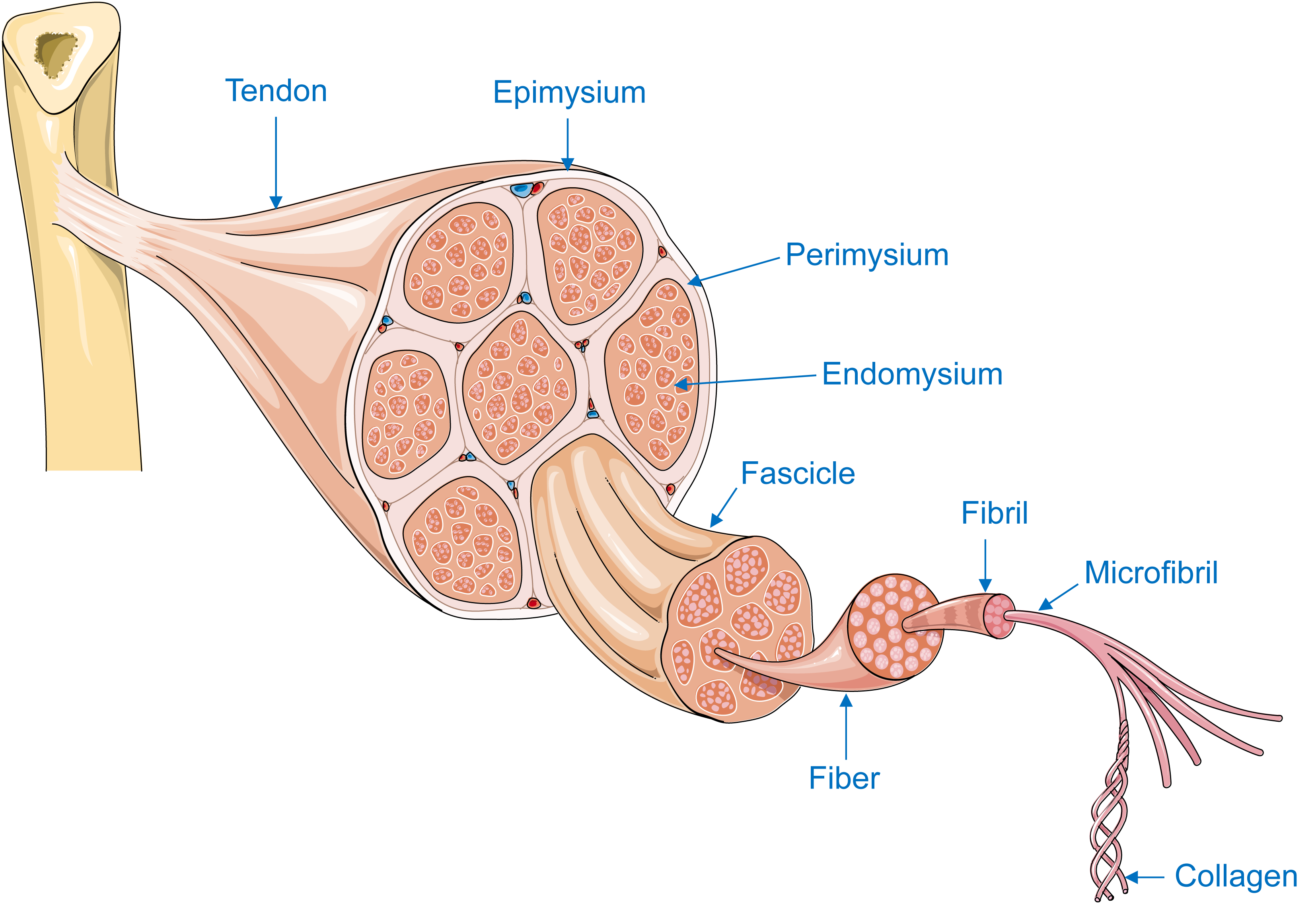
structure of skeletal muscle fiber Flashcards | Quizlet Microscopic protein filaments that make up muscle cells. Myofilaments. actin and myosin. Epimysium. covers the entire skeletal muscle. Perimysium. The connective tissue that surrounds fascicles. Endomysium. Surrounds individual muscle fibers.
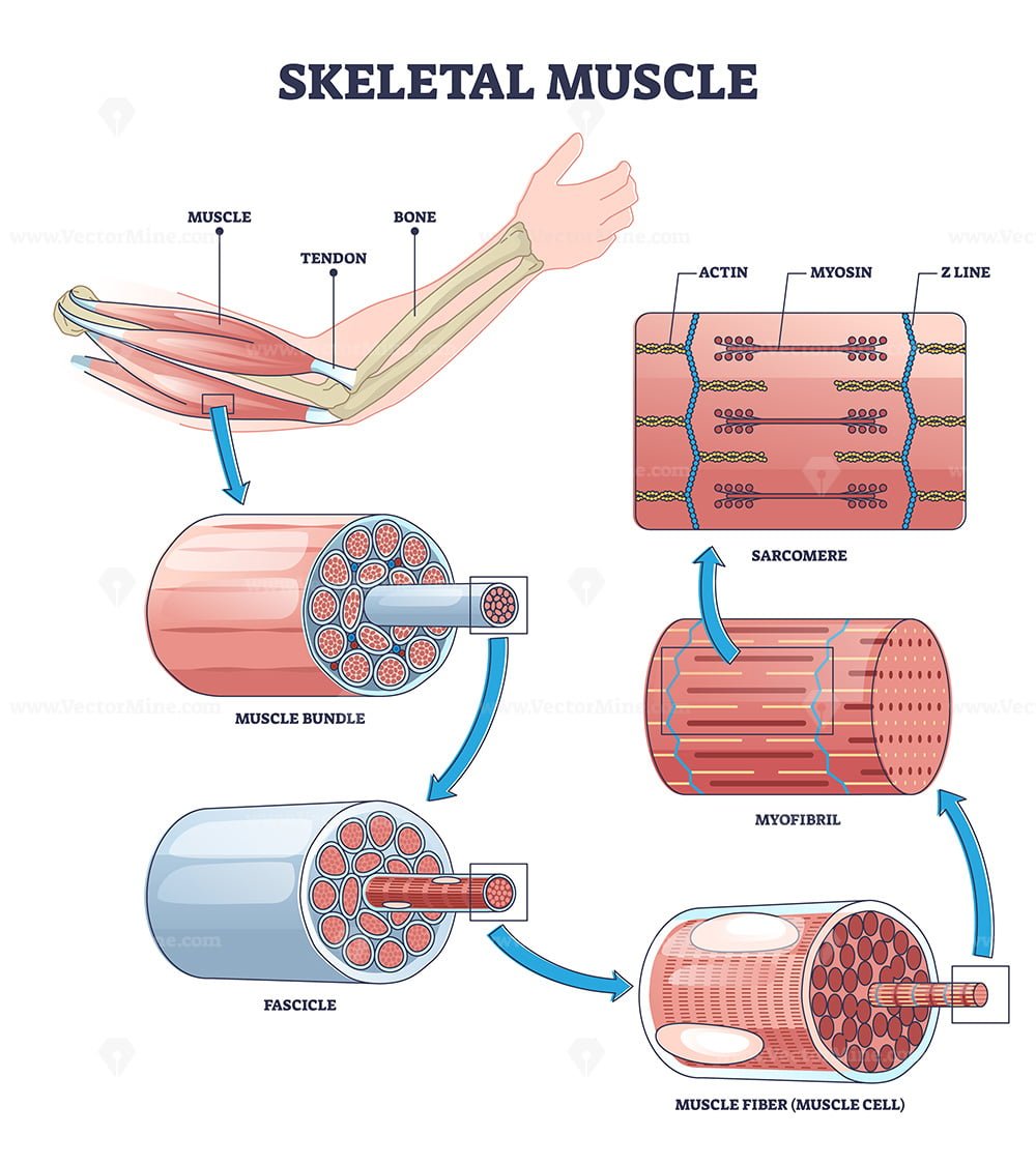
Label the structure of the muscle fiber
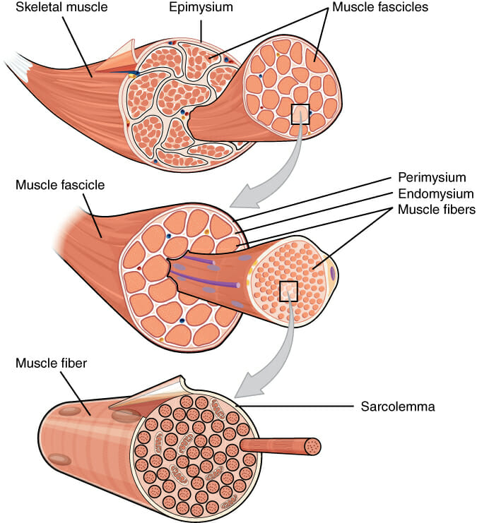
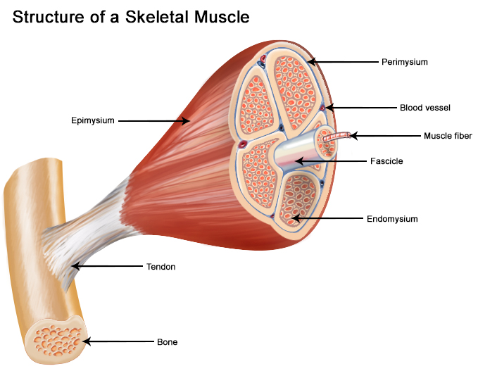
Muscles and muscle tissue: Types and functions | Kenhub There are three types of muscles. They are the: Skeletal muscles, which move bones and other structures (e.g. the eyes); Cardiac muscles, which form most of the walls of the heart and adjacent great vessels, such as the aorta; Smooth or Visceral muscles, which form part of the walls of most vessels and hollow organs, move substances through viscera such as the intestine, and control movement ... Muscle fiber label Diagram | Quizlet Start studying Muscle fiber label. Learn vocabulary, terms, and more with flashcards, games, and other study tools. Skeletal Muscle | Anatomy and Physiology I - Lumen Learning These tissues include the skeletal muscle fibers, blood vessels, nerve fibers, and connective tissue. Each skeletal muscle has three layers of connective tissue (called "mysia") that enclose it and provide structure to the muscle as a whole, and also compartmentalize the muscle fibers within the muscle (Figure 1).
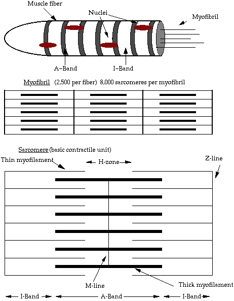
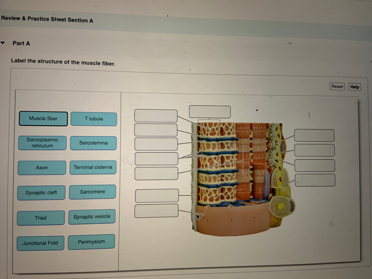
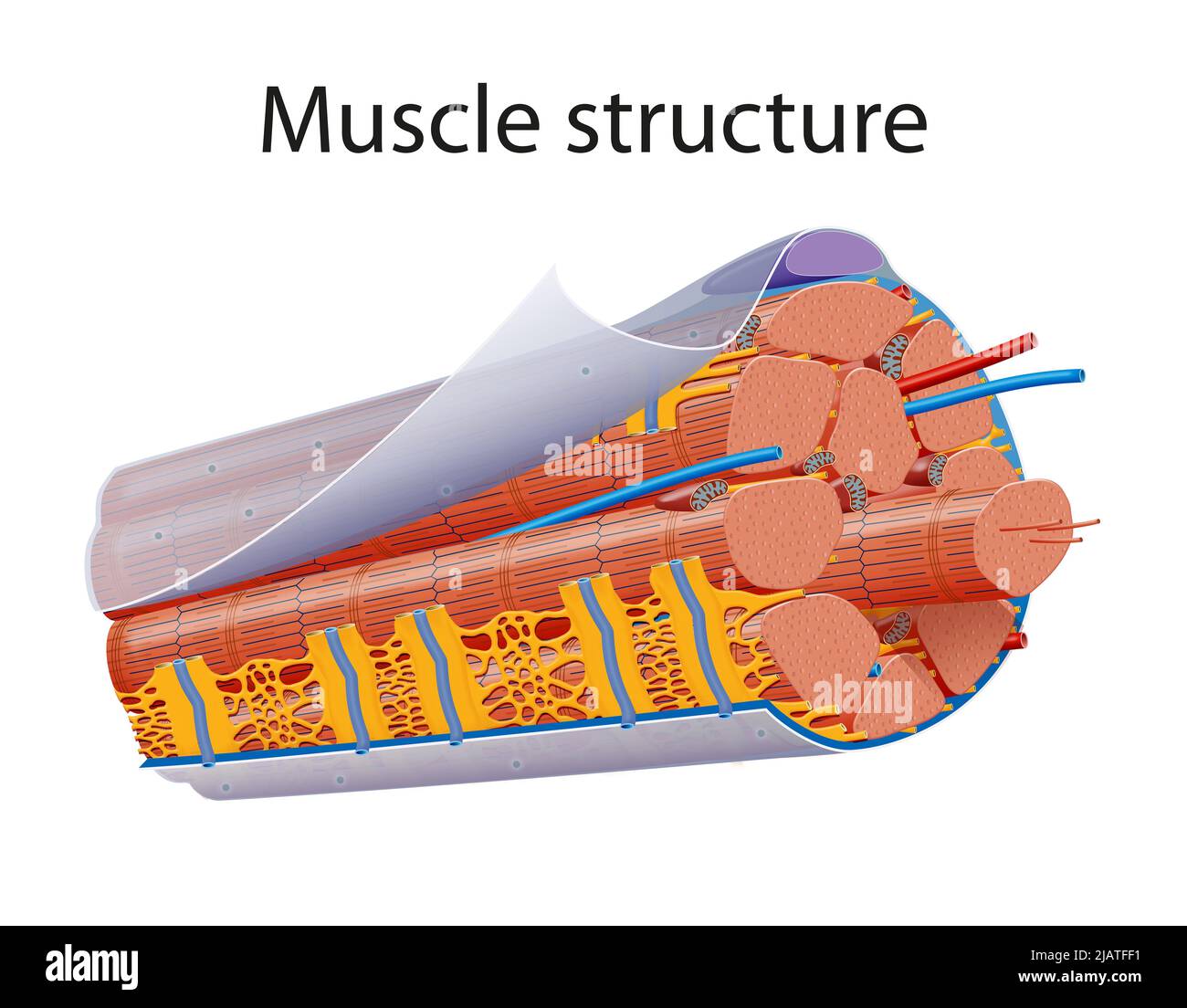
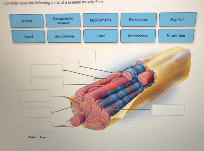
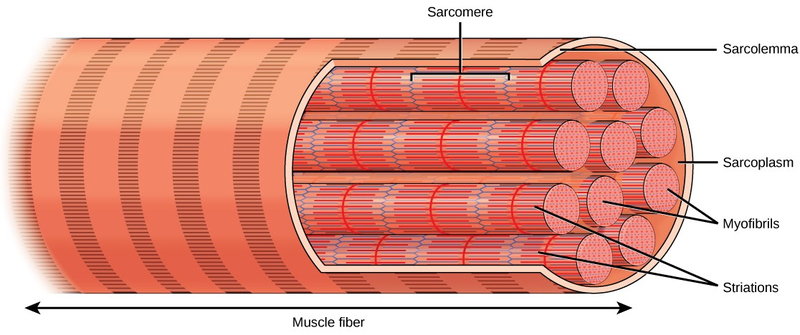
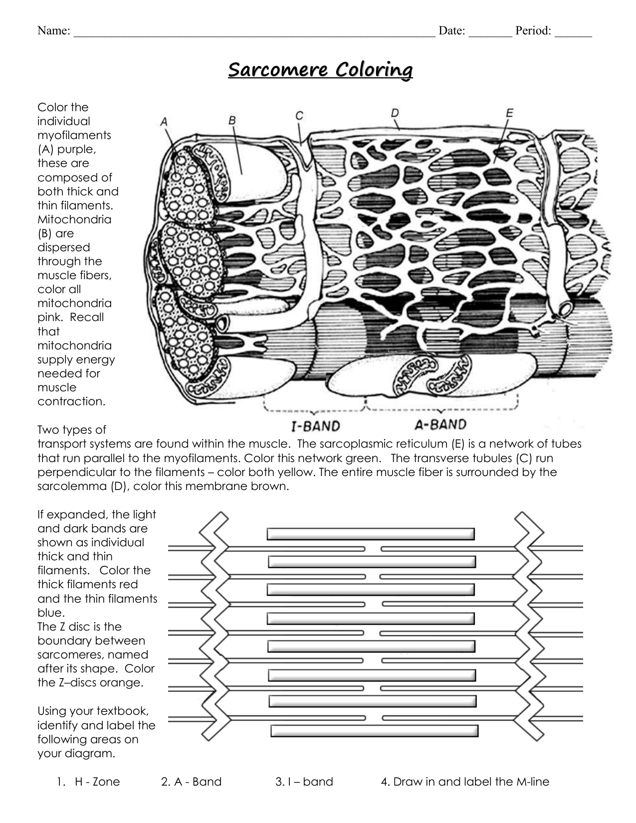
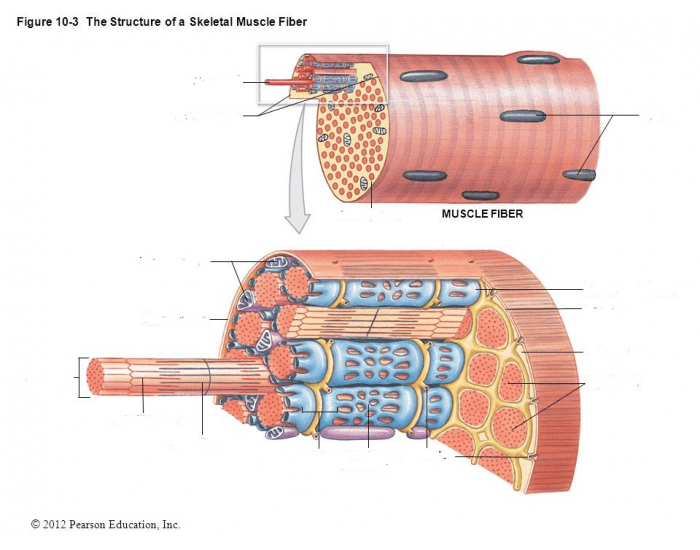
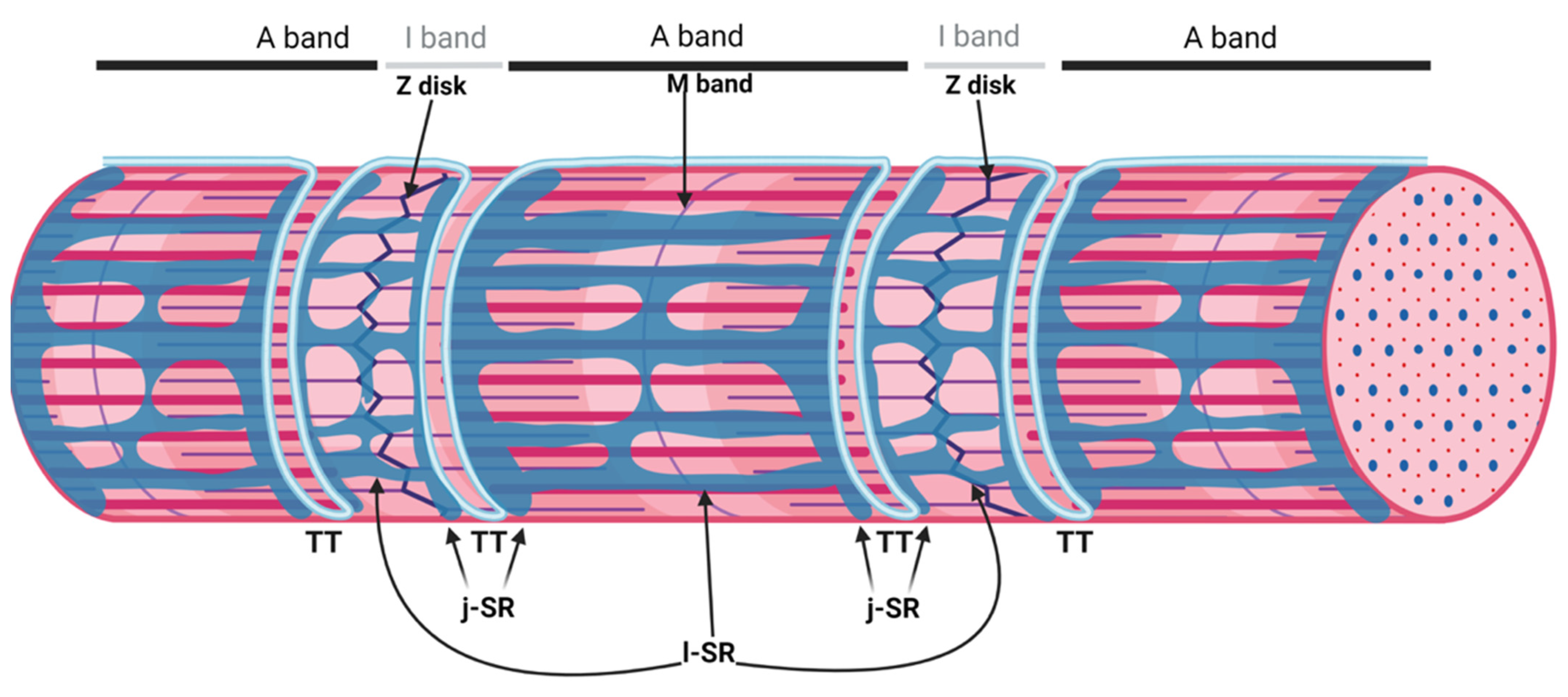
Label the structure of the muscle fiber. 10.2 Skeletal Muscle - Anatomy & Physiology Muscles attach to bones directly or through tendons or aponeuroses. Skeletal muscles maintain posture, stabilize bones and joints, control internal movement, and generate heat. Skeletal muscle fibers are long, multinucleated cells. The membrane of the cell is the sarcolemma; the cytoplasm of the cell is the sarcoplasm. Muscles Labeling - The Biology Corner Muscles Labeling. Shannan Muskopf November 11, 2020. This activity is aligned to my anatomy and physiology curriculum where students study the structure and function of muscle tissues. This has been a challenging topic to cover remotely because I can't use traditional models. Typically, I would use straws and rubber bands to model fascicles ... 11.2: Microscopic Anatomy of Skeletal Muscles - Biology LibreTexts Skeletal muscle is found attached to bones. It consists of long multinucleate fibers. The fibers run the entire length of the muscle they come from and so are usually too long to have their ends visible when viewed under the microscope. The fibers are relatively wide and very long, but unbranched. Fibers are formed from the fusion of thousands ... Solved A. Labeling Label the structure of the muscle fiber - Chegg Answer A. Label Myofibrils Sarcoplasmic reticulum Terminal cisrernae T Tubule (Transvers …. View the full answer. Transcribed image text: A. Labeling Label the structure of the muscle fiber Sarca Plasmic retiulom tencian Cistor T-woie triad Sarcomere 3. 10 4. 11 5 6 7. Sarco leme 8 9 Sin Ptis Vesiick 10.
Structure of Skeletal Muscle | SEER Training Structure of Skeletal Muscle. A whole skeletal muscle is considered an organ of the muscular system.Each organ or muscle consists of skeletal muscle tissue, connective tissue, nerve tissue, and blood or vascular tissue.. Skeletal muscles vary considerably in size, shape, and arrangement of fibers. They range from extremely tiny strands such as the stapedium muscle of the middle ear to large ... Muscle Fiber Anatomy Quiz - PurposeGames.com Muscle Fiber Anatomy — Quiz Information. This is an online quiz called Muscle Fiber Anatomy. There is a printable worksheet available for download here so you can take the quiz with pen and paper. Popular Today. 25 Cities of the USA. Cities of South West Asia. Sightseeing the US. 10.2 Skeletal Muscle - Anatomy and Physiology 2e | OpenStax Each skeletal muscle is an organ that consists of various integrated tissues. These tissues include the skeletal muscle fibers, blood vessels, nerve fibers, and connective tissue. Each skeletal muscle has three layers of connective tissue (called "mysia") that enclose it and provide structure to the muscle as a whole, and also ... Muscle Fibers: Anatomy, Function, and More - Healthline Muscle fibers can be found in skeletal, cardiac, and smooth muscles, and work to do different things in the body. ... Structure of skeletal muscle. (n.d.). training.seer.cancer.gov/anatomy ...
Solved A. Labeling Label the structure of the muscle fiber - Chegg Question: A. Labeling Label the structure of the muscle fiber 2. 10 3. 5. 6. 12 8. 10. 12 . Show transcribed image text. Expert Answer. Who are the experts? Experts are tested by Chegg as specialists in their subject area. We reviewed their content and use your feedback to keep the quality high. Muscle - Definition, Function, Types and Structure - Biology Dictionary Muscle Definition. A muscle is a group of muscle tissues which contract together to produce a force. A muscle consists of fibers of muscle cells surrounded by protective tissue, bundled together many more fibers, all surrounded in a thick protective tissue. A muscle uses ATP to contract and shorten, producing a force on the objects it is ... Muscle Fiber Labeling Quiz - PurposeGames.com This online quiz is called Muscle Fiber Labeling Quiz. It was created by member RichyT.14 and has 17 questions. ... Bone Structure (Compact Bone) by RichyT.14. 189 plays. 10p Image Quiz. Abdominal Muscle Labeling Quiz. by RichyT.14. 624 plays. 13p Image Quiz. Synovial Joint of Left Knee Labeling Quiz. Types of Muscle Tissue and Fibers | Biology for Majors II - Lumen Learning Skeletal Muscle Fiber Structure. Each skeletal muscle fiber is a skeletal muscle cell. These cells are incredibly large, with diameters of up to 100 µm and lengths of up to 30 cm. The plasma membrane of a skeletal muscle fiber is called the sarcolemma. The sarcolemma is the site of action potential conduction, which triggers muscle contraction.
Skeletal muscle structure layers with anatomical closeups ...
Skeletal Muscle | Anatomy and Physiology I - Lumen Learning These tissues include the skeletal muscle fibers, blood vessels, nerve fibers, and connective tissue. Each skeletal muscle has three layers of connective tissue (called "mysia") that enclose it and provide structure to the muscle as a whole, and also compartmentalize the muscle fibers within the muscle (Figure 1).

Interactions of Skeletal Muscles | Anatomy and Physiology I
Muscle fiber label Diagram | Quizlet Start studying Muscle fiber label. Learn vocabulary, terms, and more with flashcards, games, and other study tools.

Identify the structures of skeletal muscle. - Brainly.com
Muscles and muscle tissue: Types and functions | Kenhub There are three types of muscles. They are the: Skeletal muscles, which move bones and other structures (e.g. the eyes); Cardiac muscles, which form most of the walls of the heart and adjacent great vessels, such as the aorta; Smooth or Visceral muscles, which form part of the walls of most vessels and hollow organs, move substances through viscera such as the intestine, and control movement ...

Muscle structure – muscle under the microscope — Science ...
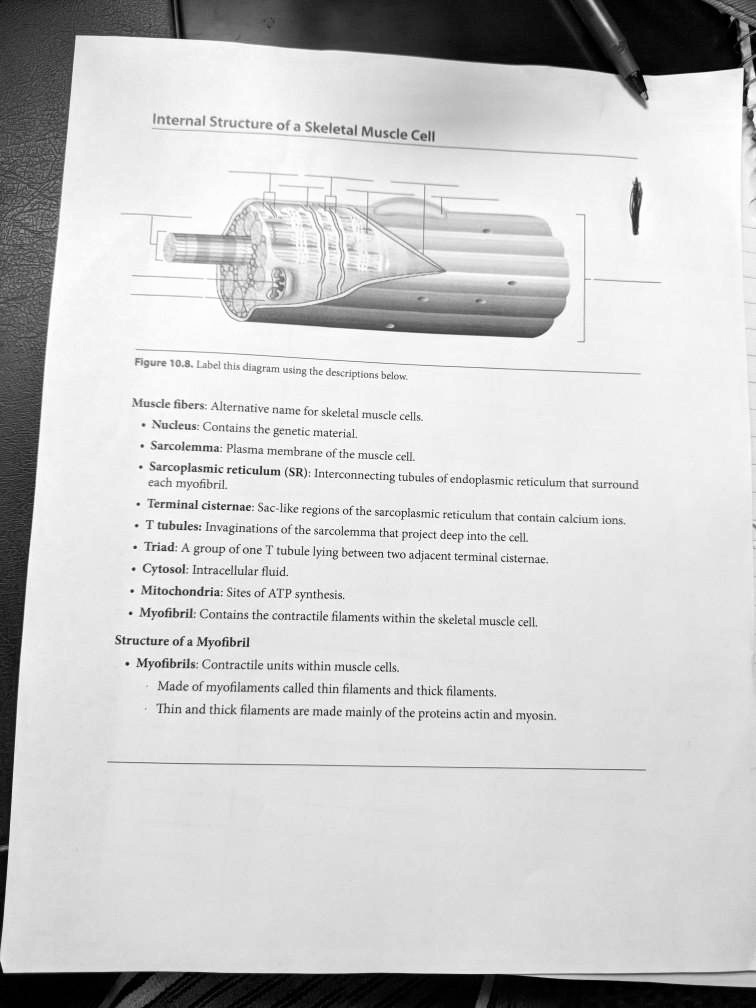
SOLVED: Internal Structure of a Skeletal Muscle Cell Figure ...

Muscle Fiber Quiz/Worksheet | Muscle, Body anatomy, Worksheets

2,600+ Skeletal Muscle Stock Photos, Pictures & Royalty-Free ...

Draw a neat diagram of the striated muscle and label the ...
SKELETAL MUSCLE ORGANIZATION

Parts of a Muscle Fiber Quiz - By ANS205_study

Neurolemmocyte On Skeletal Muscle Model - Human Anatomy ...
Muscular System Anatomy and Physiology - Nurseslabs

Skeletal Muscle Tissue Quiz for Anatomy
Structure and Composition of Muscle - Meat Science
Answered: Label the structure of the muscle fiber | bartleby

loadBinary_011.gif
Muscle - Definition, Function, Types and Structure | Biology ...

Lab Practical #2 Flashcards | Quizlet
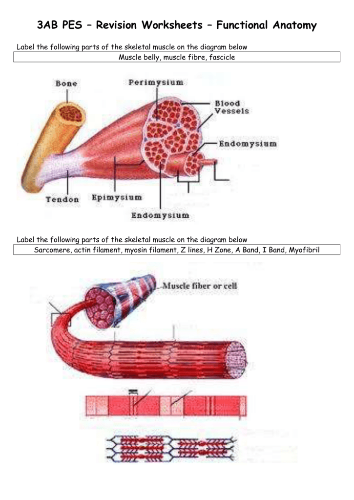
3AB PES – Revision Worksheets – Functional - PE Studies

Sliding Filament Theory: Steps for Muscle Contraction
Solved Bone Fascia (located between adjacent muscles) | Chegg.com
A test of strength for artificial muscles | Feature ...
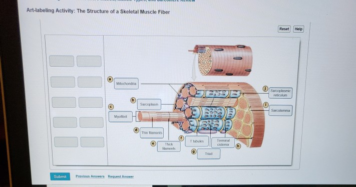
Solved Art-labeling Activity: The Structure of a Skeletal ...

Diagram of Skeletal Muscle
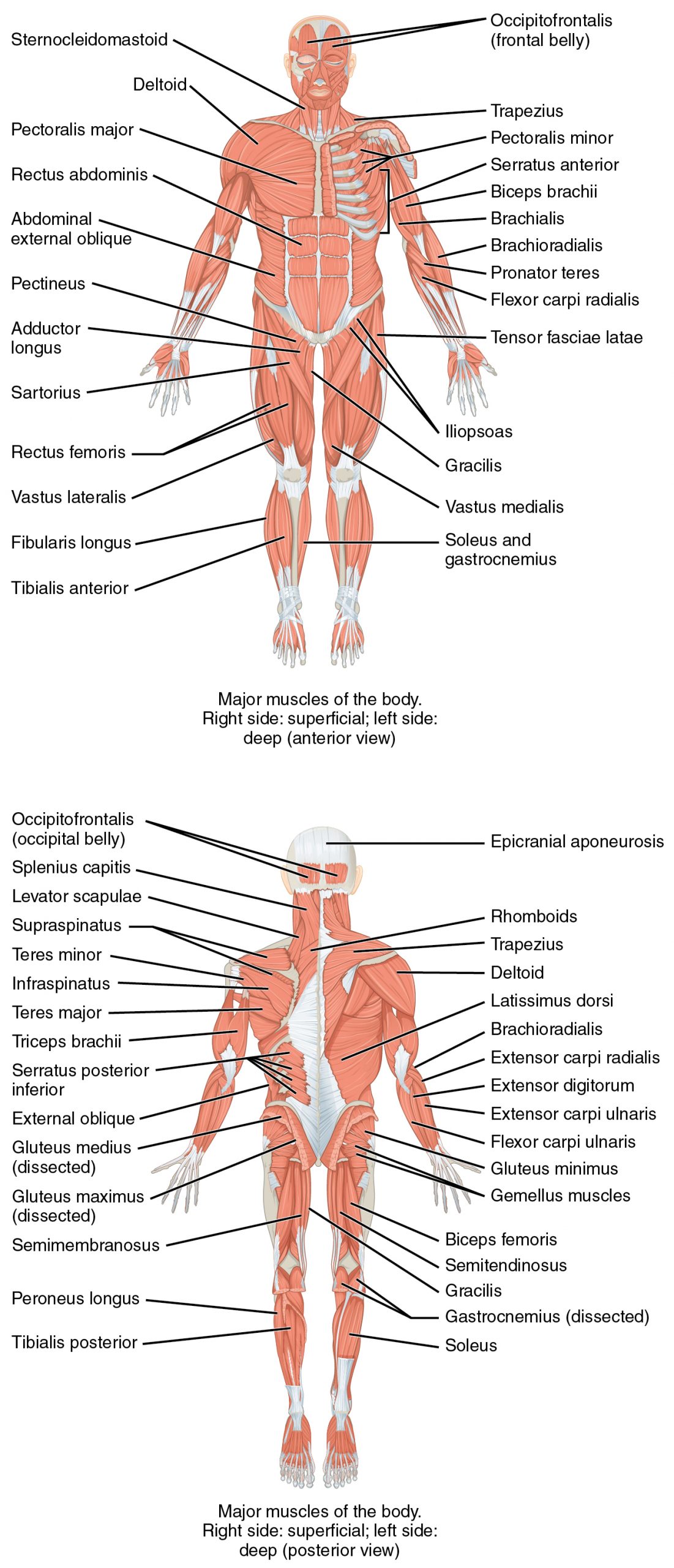
11.3 Explain the criteria used to name skeletal muscles ...
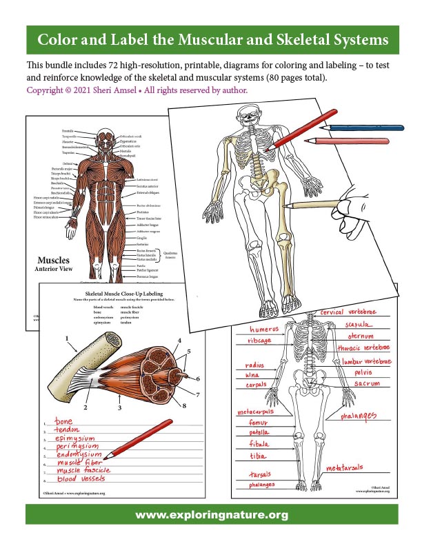
Color and Label the Muscular and Skeletal Systems ...
Servier - Drawing Structure of the skeletal muscle - English ...

Label the following in a diagram of a skeletal muscle fiber ...

Muscular System Objectives: A.Describe the purpose of the ...
PRACTICE SHEET FOR JOINT, SKELETAL MUSCLE STRUCTURE, TYPES OF ...
Leg muscle fiber hi-res stock photography and images - Alamy

BIO 200 Chapter 9 - Muscle Tissue Physiology Flashcards | Quizlet
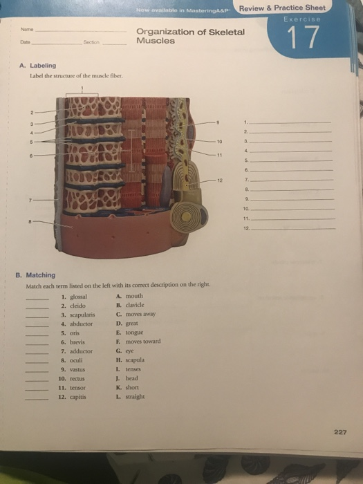
Solved Now available in MasteringA&P Review & Practice Sheet ...

The Skeletal Muscle Fiber Stock Illustration - Download Image ...
Solved Correctly label the following parts of a skeletal ...
Skeletal Muscle Fiber Structure and Function | Open Textbooks ...
SEER Training: Structure of Skeletal Muscle
Sarcomere and Sliding filament Theory Coloring Worksheet

Myofibril - Wikipedia
Structure of a skeletal muscle fiber Quiz
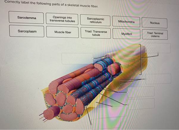
Solved) - Correctly label the following parts of a skeletal ...

Lab 17: Skeletal Muscle Structure
Biomolecules | Free Full-Text | The Sarcoplasmic Reticulum of ...
Post a Comment for "43 label the structure of the muscle fiber"